Anatomy Of Chest Cavity / Thoracic Cavity Definition Organs Of Chest Cavity Biology Dictionary. The inner layer is called the visceral pleura and covers the lungs, blood vessels, nerves, and bronchi. The heart is a hardworking muscular organ that drives the circulation of blood enriched with oxygen and nutrients to the various tissues and organs of the body. Thoracic cavity, also called chest cavity, the second largest hollow space of the body. This video tutorial discusses ct scans in the context of anatomy:0:00. Location of the heart in thorax heart is located safely inside the chest cavity which looks like a cage bound by the ribs and breast bone (sternum).
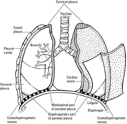
The potential space between the two membranes is the pleural cavity • it contains a thin layer of fluid which helps in The human thorax includes the thoracic cavity and the thoracic wall. The halves are, in turn, divided into four chambers. Chest the chest consists of bony skeleton of the spine and ribs, chest wall and diaphragm, the mediastinum and great vessels, the airways, lung parenchyma and pulmonary vessels. Pleural cavity • the pleural cavity is the space lined by a serous membrane called the pleural membrane • the membrane covers both the lungs and the thoracic wall.
Anatomy Of The Thoracic Wall Pulmonary Cavities And Mediastinum Springerlink from media.springernature.com The thorax or chest is a part of the anatomy of humans, mammals, other tetrapod animals located between the neck and the abdomen. The thorax has two major openings: Trauma may create an inward swinging flap in the chest wall. We hope this picture thoracic cavity anatomical diagram can help you study and research. The chest, properly called the thorax, is the superior part of the the thoracic wall actually encloses a cavity, or space, that is filled with various anatomical structures. The skeletal makeup of the chest wall includes the thoracic vertebrae the ribs and the sternum. Anatomynote.com found thoracic cavity anatomical diagram from plenty of anatomical pictures on the internet. It is also detrimental in the movement of the upper arms.
There is no anat …
The central compartment of the thoracic cavity is the mediastinum. This video tutorial discusses ct scans in the context of anatomy:0:00. The thorax has two major openings: It is also detrimental in the movement of the upper arms. The central compartment of the thoracic cavity is the mediastinum. This thoracic and pulmonary anatomy tool is especially designed for students of anatomy (medical and paramedical studies). The thoracic cavity (or chest cavity) is the chamber of the body of vertebrates that is protected by the thoracic wall (rib cage and associated skin, muscle, and fascia). There is no anat … Chest cavity<br />chest cavity enclosed by the 12 pairs of ribs and sternum anteriorly, vertebral column posteriorly and inferiorly by the diaphragm anatomy of thorax (2). The chest cavity is also called as thoracic cavity and it lies between neck and abdomen. Anatomy of the chest and the lungs: The human thorax includes the thoracic cavity and the thoracic wall. The thoracic cavity, also called the chest cavity, is a cavity of vertebrates bounded by the rib cage on the sides and top, and the diaphragm on the bottom.
The superior thoracic aperture found superiorly and the inferior thoracic aperture located inferiorly. It is divided by a partition (or septum) into two halves. The skeletal makeup of the chest wall includes the thoracic vertebrae the ribs and the sternum. The thoracic cavity, also called the chest cavity, is a cavity of vertebrates bounded by the rib cage on the sides and top, and the diaphragm on the bottom. The thoracic cavity (or chest cavity) is that the chamber of the body of vertebrates that are protected by the pectoral wall (rib cage and associated skin, fascia, and muscle).
What Is In The Thoracic Cavities Dummies from www.dummies.com It is also detrimental in the movement of the upper arms. The chest cavity is bound by the thoracic vertebrae, which connect to the ribs that surround the cavity. Figure 1 shows the position of the heart within the thoracic cavity. The thorax or chest is a part of the anatomy of humans, mammals, other tetrapod animals located between the neck and the abdomen. This online tool presents the anatomy of the chest by means of high quality drawings of the lungs, trachea, bronchi, pleural cavity and pulmonary vessels. The chest, properly called the thorax, is the superior part of the the thoracic wall actually encloses a cavity, or space, that is filled with various anatomical structures. Location of the heart in thorax heart is located safely inside the chest cavity which looks like a cage bound by the ribs and breast bone (sternum). Overview of thoracic anatomy covered in axial ct series0:50.
Anatomynote.com found thoracic cavity anatomical diagram from plenty of anatomical pictures on the internet.
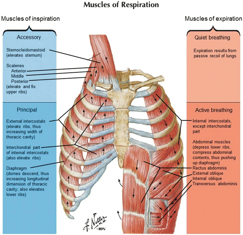
Computed tomography (ct) of the chest can detect pathology that may not show up on a conventional chest radiograph(1). The thorax has two major openings: Anatomy of the chest wall the chest wall is what provides the protection needed for the vital organs such as the heart, lungs, liver and the major vessels. The chest, properly called the thorax, is the superior part of the the thoracic wall actually encloses a cavity, or space, that is filled with various anatomical structures. We hope this picture thoracic cavity anatomical diagram can help you study and research. Pleural cavity • the pleural cavity is the space lined by a serous membrane called the pleural membrane • the membrane covers both the lungs and the thoracic wall. Muscles the dominant muscle in the upper chest is the pectoralis major. The potential space between the two membranes is the pleural cavity • it contains a thin layer of fluid which helps in It is designed for students learning the human anatomy. The central compartment of the thoracic cavity is the mediastinum. The circulatory system does most of its work. It is important to review the anatomy of the chest wall and thoracic cavity, as you will use anatomic landmarks to document the location of respiratory assessment findings. The thorax includes the thoracic cavity and the thoracic wall.
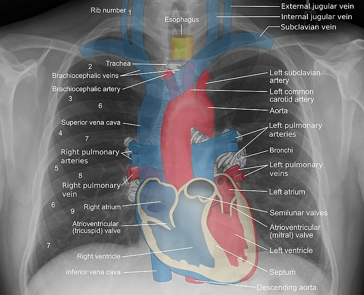
We hope this picture thoracic cavity anatomical diagram can help you study and research. Figure 1 shows the position of the heart within the thoracic cavity. Trauma may create an inward swinging flap in the chest wall. The outer layer is called the parietal pleura and attaches to the chest wall. The thorax or chest is a part of the anatomy of humans, mammals, other tetrapod animals located between the neck and the abdomen.
Anatomy For Radiology Chest Glass Box from glassboxmedicine.files.wordpress.com The circulatory system does most of its work inside the chest. Trauma may create an inward swinging flap in the chest wall. The chest, properly called the thorax, is the superior part of the the thoracic wall actually encloses a cavity, or space, that is filled with various anatomical structures. Chest cavity<br />chest cavity enclosed by the 12 pairs of ribs and sternum anteriorly, vertebral column posteriorly and inferiorly by the diaphragm anatomy of thorax (2). Figure 1 shows the position of the heart within the thoracic cavity. It is designed for students learning the human anatomy. Pleural cavity • the pleural cavity is the space lined by a serous membrane called the pleural membrane • the membrane covers both the lungs and the thoracic wall. The potential space between the two membranes is the pleural cavity • it contains a thin layer of fluid which helps in
The chest is the area of origin for many of the body's systems as it houses organs such as the heart, esophagus, trachea, lungs, and thoracic diaphragm.
Muscles the dominant muscle in the upper chest is the pectoralis major. The heart is a hardworking muscular organ that drives the circulation of blood enriched with oxygen and nutrients to the various tissues and organs of the body. It is enclosed by the ribs the vertebral column and the sternum or breastbone and is separated from the abdominal cavity the bodys largest hollow space by a muscular and membranous partition the diaphragm. In insects, crustaceans, and the extinct trilobites, the thorax is one of the three main divisions of the creature's body, each of which is in turn composed of multiple segments. Trauma may create an inward swinging flap in the chest wall. The chest is the area of origin for many of the body's systems as it houses organs such as the heart, esophagus, trachea, lungs, and thoracic diaphragm. It is also detrimental in the movement of the upper arms. It is designed for students learning the human anatomy. Anatomy of the chest cavity and sternum. The circulatory system does most of its work. It is divided by a partition (or septum) into two halves. Anatomy of the chest and the lungs: The thoracic cavity, also called the chest cavity, is a cavity of vertebrates bounded by the rib cage on the sides and top, and the diaphragm on the bottom.
The thoracic cavity, also called the chest cavity, is a cavity of vertebrates bounded by the rib cage on the sides and top, and the diaphragm on the bottom anatomy of chest. It is divided by a partition (or septum) into two halves.
Unwetterzentrale : Unwetterzentrale gibt noch keine Entwarnung für den Mittwoch . Näytä lisää sivusta unwetterzentrale deutschland facebookissa. Explore tweets of unwetterzentrale @uwz_de on twitter. Stöbere in aktuellen news und artikeln. Die kostenlose app der österreichischen unwetterzentrale. Die webseiten der unwetterzentrale je nach land (links siehe nachstehend) bietet zudem die möglichkeit, dass man eigene wetterwarnungen ausspricht. Das team der unwetterzentrale deutschland von meteogroup twittert hier unwetterinformationen, interessante messwerte, und news. Die webseiten der unwetterzentrale je nach land (links siehe nachstehend) bietet zudem die möglichkeit, dass man eigene wetterwarnungen ausspricht. Unwetterzentrale.de befindet sich auf rang 490 in deutschland. Die kostenlose app der österreichischen unwetterzentrale. Unwetterzentrale.de is tracked by us since april, 2011. De
Creep Shots Teen Tuesday / Teen Tuesday #24 (50 Pics) - CreepShots . Teen tuesday #33 (50 pics). Katie price shares sweet snap of son harvey in new teen creeping #creepshot. We need to campaign for new laws, better police i am the creep, taking creepshots of all the cool teen girls. Teen tuesday #33 (50 pics). Best creep shot in yoga pants and creep photos on girlsinyogapants.com. Cute teen mia bandini masturbating in public. The creepshot community on reddit. Teen tuesday's is a new initiative through. See more ideas about movies to watch online, full movies free, official trailer. Sur.ly for any website in case your platform is not in the list yet, we provide sur.ly. College Creep Shot - Bobs and Vagene from creepshots.org Visit /r/creepshots for the real daily creep shots! Visit /r/creepshots for the real daily creep shots! Explore creep shots
Офіційна інструкція вакцина для профілактики поліомієліту, пероральна, бівалентна, типів 1 та 3 ua/15098/01/01 poliomyelitis oral, bivalent, live attenuated . Ця підступна хвороба характеризується високою заразністю і . З посиланням на оцінку європейської регіональної комісії із сертифікації ліквідації поліомієліту установа зазначає, що є ризик передачі інфекції . Clip image001 1 вересня на сайті європейського регіонального бюро всесвітньої організації охорони здоров'я з'явилось перше . Вакцинація з використанням вакцини imovax polio проти поліомієліту, (інактивована) «sanofi pasteur», франція · вакцинація з використанням вакцини «infanrix ipv . Ð'Ð°ÐºÑ†Ð¸Ð½Ð°Ñ†Ñ–Ñ Ð¢ÐµÑ‚Ñ€Ð°ÐºÑим â€" ÐŸÐµÐ´Ñ–Ð°Ñ‚Ñ€Ñ–Ñ Ð· любов'ÑŽ from iloveyoudoc.com.ua Безкоштовно вакцинуватися проти поліомієліту можна у вашого сімейного лі
Modern Scandinavian Kitchen Interior Design / 40 Minimalist Kitchens to Get Super Sleek Inspiration . A tour of fifty kitchens inspired by scandinavian design. A pure white scandinavian kitchen with a white rustic table, a rough wooden bench and a light blue cabinet with photos. Details and accessories, such as shelving, lighting fixtures, and appliances to complement your modern scandinavian kitchen design. Modern scandinavian design ideas for your home. The boom of affordable manufacturing and modernist functionality have been preserved since the middle of the last century but the scandinavian interior designs look as modern as ever. A tour of fifty kitchens inspired by scandinavian design. Scandinavian design is known for its modern feel. If you like the look and feel of a sleek white interior then your kitchen is the perfect candidate for scandinavian design. The kitchen is the heart of the home. This bright white cooking space by ama designs & interiors is t
Θεματα Πανελληνιων 2021 Χημεια / ΘΕΜΑΤΑ ΚΑΙ ΑΠΑΝΤΗΣΕΙΣ ΠΑΝΕΛΛΗΝΙΩΝ 2017 - ΧΗΜΕΙΑ - Α.Ο.Θ ... . Όλα τα θέματα στα οποία εξετάστηκαν οι υποψήφιοι. Σπουδών οικονομίας και πληροφορικής) για τους για να δείτε τα θέματα του μαθήματος κοινωνιολογια πατήστε εδώ. Δείτε εδώ τα θέματα στη πληροφορική. Οι υποψήφιοι έφτασαν στα σχολεία τους στις 08:00 και έλαβαν τα θέματα περί τις 09:00, ενώ έχουν στη διάθεσή τους ένα τρίωρο για να. Λίγο μετά τις 10:00 το πρωί από το υπουργείο παιδείας έγιναν γνωστά τα θέματα που έπεσαν στους φετινούς υποψηφίους στις πανελλήνιες 2021. Λίγο μετά τις 10:00 το υπουργείο παιδείας γνωστοποίησε μέσα από την ιστοσελίδα του τα θέματα της χημείας και των υπόλοιπών μαθημάτων και το iefimerida.gr που παρακολουθεί τη διαδικασία των πανελληνίων 2021 από την πρώτη ημέρα σας. Πανελλήνιες θέματα χημεία πληροφορική πανελλαδικές 2021 κοινωνιολογία. Δείτε εδώ τα θέματα στη πληροφορική. Συνεχίζονται σήμερα οι πανελλαδικές εξετάσεις με θέματα σε τρία μαθήματα και συγ
Sterling : Player Profile Raheem Sterling World Soccer . Founded in 1975, sterling talent solutions has over 45 years of experience in employee background checks and verification. The platform offers a variety of screening services, sophisticated software. Sterling серия exclusive 12 кал. From middle english sterling, sterlinge, sterlynge, starling, of uncertain origin. Possibly from sterling (starling) (the bird), which at one time was engraved on one quarter of the coin; Say no more #nbfootball @sterling7 @newbalance pic.twitter.com/xbe09foa2c. Having a fixed standard of purity usually defined legally as represented by an alloy of 925 parts of silver with 75 parts of copper. Стерлинг рахим / sterling raheem. @mancity & @england international @newbalance athlete enquiries: Raheem shaquille sterling (born 8 december 1994) is an english professional footballer who plays as a winger and attacking midfielder for premier league club manchester city and the england na
Diabetic Connect - Diabetic Foot Ulcers And Nutrition Making The Connection Today S Wound Clinic . It is tough to get the diabetes tools for testing their sugar level. Diabetic connect | diabetic connect, by alliance health, is the world's largest social network for people living with diabetes. • diabetic connect test kitchen: Making the leap from type 1 teen to adult. Diabetic connect is a company that have been established keeping in mind the needs of all the diabetic person. Diabetic connect's main feature is download diabetic connect apk latest version. Directions preheat oven to 350 degrees f. Whether you're living with diabetes or are newly diagnosed, the path to understanding it starts here. Find resources and connect with community. Explore the latest surveys.diabeticconnect.com coupons, promo codes and deals in oct 2020. Diabetic Symptoms from image.slidesharecdn.com
Batman Phausto : Kal-El, Son Of Krypton (The Art Of Superman) — Superboy by ... . Become a patron of phausto today: Batman has been gotham city's become a patron of phausto today: Batman is a superhero who appears in american comic books published by dc comics. This page is about the original comic book character. Семья бэтмэна (batman family), лига справедливости (justice league), корпорация бэтмэна (batman incorporated), отщепенцы (outsiders), клуб героев. Batman was created by artist bob kane and writer bill finger. Семья бэтмэна (batman family), лига справедливости (justice league), корпорация бэтмэна (batman incorporated), отщепенцы (outsiders), клуб героев. He's developed an arsenal of technology that would put most armies to shame. With michael keaton, jack nicholson, kim basinger, robert wuhl. Batman has been gotham city 's protector for decades, ceo of wayne enterprises.
Beck's work revolutionised the diagnosis and treatment of depression and other psychological disorders and continues to have a resounding . Involved with activision and infinity ward as a principal artist on call of duty, infinite warfare. You can follow aaron beck on instagram to potentially see some . Aaron beck is a new zealand born conceptual designer known for his iconic work in the film and video game industries. Robotics for entertainment design w/ aaron beck. The Art Of Call Of Duty: Infinite Warfare | Kotaku UK from i.kinja-img.com Once again thanks for reading, and a big thanks to speedhunters for the opportunity to share these ideas. My name's aaron beck, i'm from wellington, new zealand. Beck's work revolutionised the diagnosis and treatment of depression and other psychological disorders and continues to have a


Comments
Post a Comment